
Negative Staining
Many organic or biological samples are mainly composed oflight elements such as carbon (C), hydrogen (H), and oxygen (O). During Transmission Electron Microscopy (TEM)The electron scattering ability is relatively weak during observation, so the image contrast is usually low, making it difficult to clearly identify its microstructure. In order to enhance the image recognition and observation effect, it is often utilizedheavy metal dyeStaining the background around the sample to create a clear contrast of the sample outline in electron microscope images is calledNegative Staining。
The principle of negative staining technology is to use high electron density metal stains (such as phosphotungstic acid, uranyl acetate, etc.) to coat the sample, forming a dark background, while the sample itself presents a brighter image outline, allowing its morphology and structure to be clearly displayed in TEM images. Through this method, rapid observation can be made.virus particles, protein complexes, nano drug carriers, liposomes, and various biomedical nanomaterialsThe shape and size distribution.
Due to the metal negative staining havingThe sample preparation process is simple, the observation efficiency is high, and it can effectively enhance the image contrast.Due to its advantages, it is widely used inBiomedical research, nanomedicine development, virus structure observation, and biological nanomaterials analysisIn the same field. The TEM images after negative staining can assist researchers in quickly grasping the morphology of nanostructures and the distribution of particles, providing important microstructural information for the development and quality assessment of biomedical materials.
-
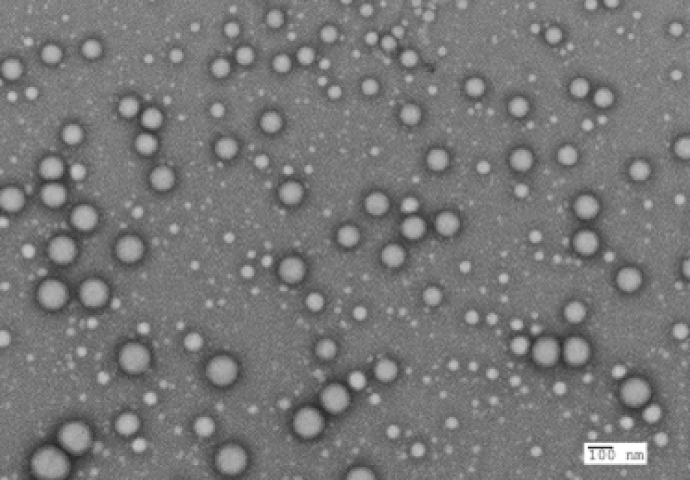 Negative staining results of PLGA particles
 -
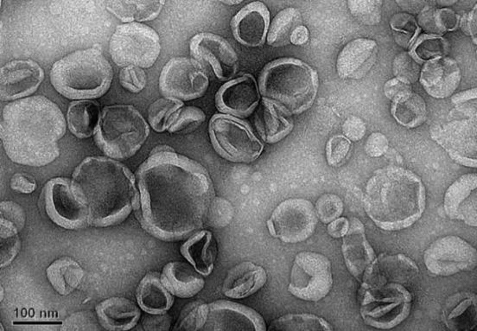 Negative staining results of Liposome particles