
For high water content biological samples such as cells, bacteria, biological tissues, and organ specimensand some biomedical materials, when conductingTransmission Electron Microscopy (TEM)Before observation, a complete sample preparation process is usually required. Common processing steps includeGlutaraldehyde Fixation, Dehydration, Resin Embedding, and UltramicrotomyThe same procedures are followed to ensure that the samples can maintain their original microscopic structure in the high vacuum environment of the electron microscope.
After completing the resin embedding, the sample can be usedUltramicrotomePrepare ultra-thin sections with a thickness of about tens of nanometers, and then collect the samples inCopper mesh (TEM Grid)Observation is conducted. Through this sample preparation technique, the resolution and structural recognition of electron microscope images can be effectively improved, allowing researchers to clearly observe the internal structures of cells, the morphology of bacteria, and the microscopic structural features of biological tissues.
General biological samples such as cells, bacteria, biological tissues and medical materialsetc., can all be achieved through appropriate sample pre-treatment andRoom temperature or frozen ultrathin section technologysample preparation and high-resolution structural imaging observation in transmission electron microscopy to obtain nanoscale microscopic structural information. This technology is widely used inBiomedical materials research, cell structure analysis, virus and nanodrug observation, and medical device material analysis. and other fields.
-
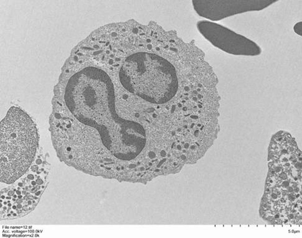 White blood cell slice image
 -
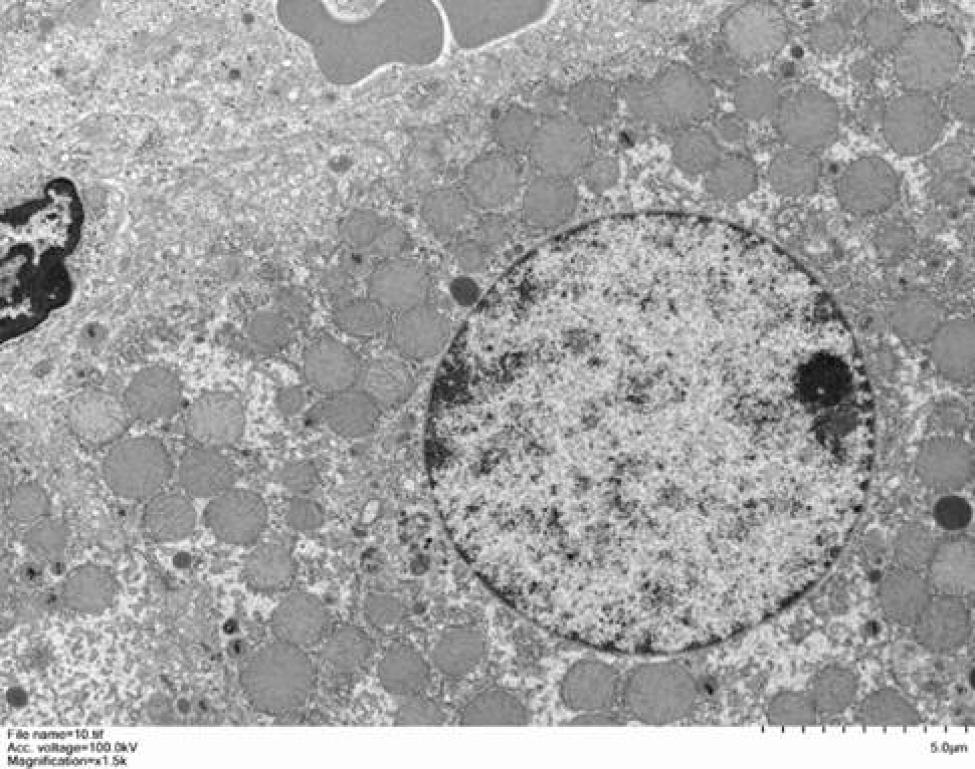 Mouse liver tissue section image